
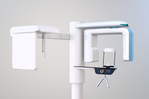
Modern endodontists take full advantage of the latest advancements in order to enhance the care that they’re able to offer their patients. 3D imaging is a key example of this; this state-of-the-art technology has proven to be an invaluable tool for diagnosing oral health issues and planning complex endodontic procedures. Below is a closer look at how 3D imaging works and the important benefits that it offers.
What is 3D Imaging?
3D imaging is sometimes also referred to as cone-beam computed tomography, or CBCT for short. The technology can be used to create a three-dimensional model of your mouth that shows the teeth, the jawbone, and various oral structures. Thanks to this model, your endodontist can get a comprehensive picture of your oral landscape, allowing them to plan your treatment down to the smallest detail.
How Exactly Does 3D Imaging Work?
The 3D imaging process involves a CBCT scanner that emits a cone-shaped X-ray beam. As the scanner rotates around your head, it takes hundreds of pictures of your mouth and jaw. Then, with the help of special software, all of the captured pictures are combined to create a three-dimensional model of your mouth that can be viewed from any angle.
How Do Endodontists Use 3D Imaging?
With 3D imaging, endodontists can get a detailed look at the internal anatomy of your teeth. Not only can the technology help identify any abnormalities that need to be accounted for, but it can also reveal the location of hidden canals. This information can make a significant difference when planning root canal treatments and other endodontic procedures. On top of that, 3D imaging can be used to determine whether the mouth is healing properly after treatment.
What are the Advantages of 3D Imaging?
There are several reasons why so many endodontists nowadays utilize 3D imaging:
- More Accurate Diagnoses: 3D imaging can capture details that might not show up in a traditional two-dimensional X-ray. This allows your endodontist to assess your oral health issues as accurately as possible so that they can take the appropriate steps to address them.
- Improved Treatment Plans: 3D imaging can provide vital information about the anatomy of your teeth. Learning these details ahead of time will make it easier for your endodontist to plan your treatment and ensure that the inside of the tooth is cleaned as thoroughly as possible.
- Less Radiation: CBCT scanners are designed to minimize the amount of radiation that patients are exposed to. As such, the technology is considered to be very safe.
- No Discomfort: There’s no need to be worried about having 3D imaging performed; the process is painless and can typically be completed in less than a minute.
In short, 3D imaging has become an important part of modern endodontic care. By using it to carefully plan root canals and other procedures, your endodontist can maximize the chances of your treatment being successful while keeping risks to a minimum. It’s just one example of how advancements in technology continue to transform the field of endodontics.
About the Author
Dr. Tam Trinh earned his dental degree at the University of Illinois Chicago College of Dentistry, and he completed his endodontic residency at the University of North Carolina at Chapel Hill. He is a member of the American Association of Endodontists and various other organizations. His practice, MeTa Endodontics, takes full advantage of 3D scanning and other technological advancements to bring patients high-quality care. If you need the services of a knowledgeable endodontist in Deerfield, you can schedule an appointment with Dr. Trinh by visiting his website or calling (847) 920-6382.